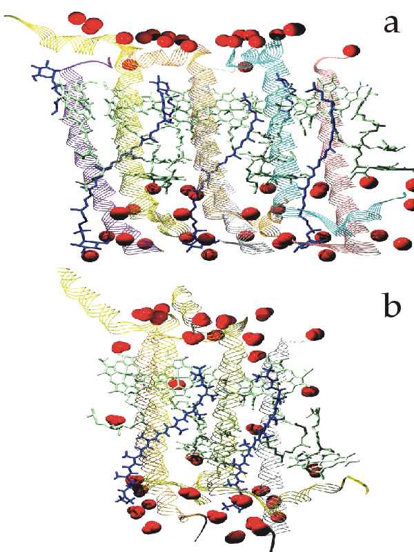
 Figure B-7_a&b
LH-II of Rhodopseudomonas acidophila & Rhodospirillum molischianum.
Figure B-7_a&b
LH-II of Rhodopseudomonas acidophila & Rhodospirillum molischianum.
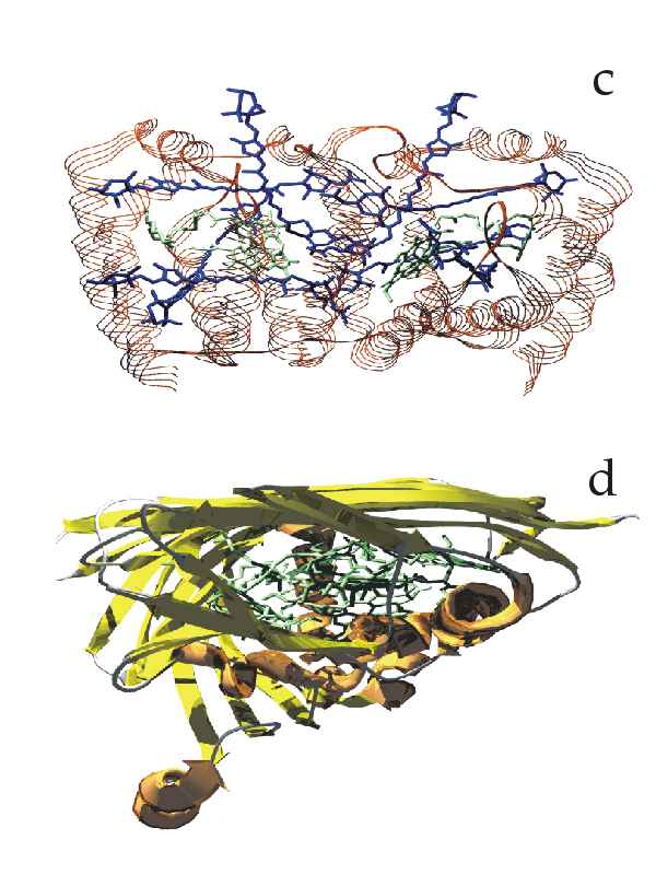 Figure B-7_c&d
PCP of Amphidinium carterae & bacteriochlorophyll protein of Prosthecochloris aestuarii.
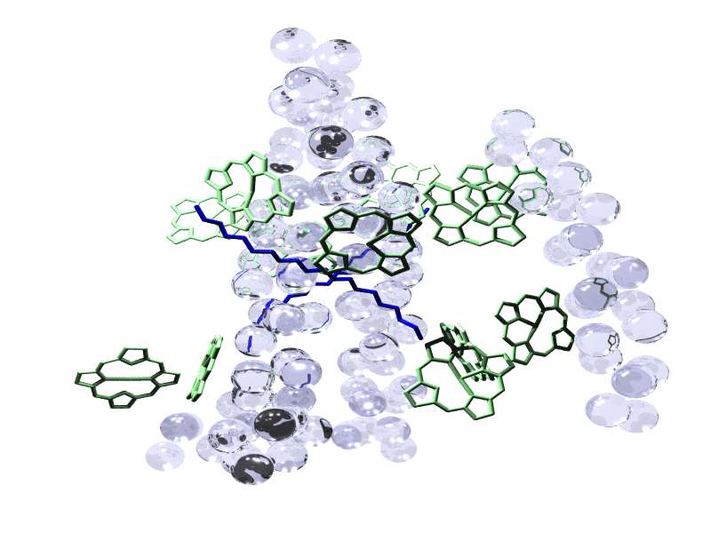 C-alfa_only
initial structure.
C-alfa_only
initial structure.
This image didn't quite make it in the thesis. It
shows the initial structure I received and had to work on. The
initial information was just the coordinates of the C-alfa atoms,
represented by the spheres in the image, and of the chlorophyll rings.
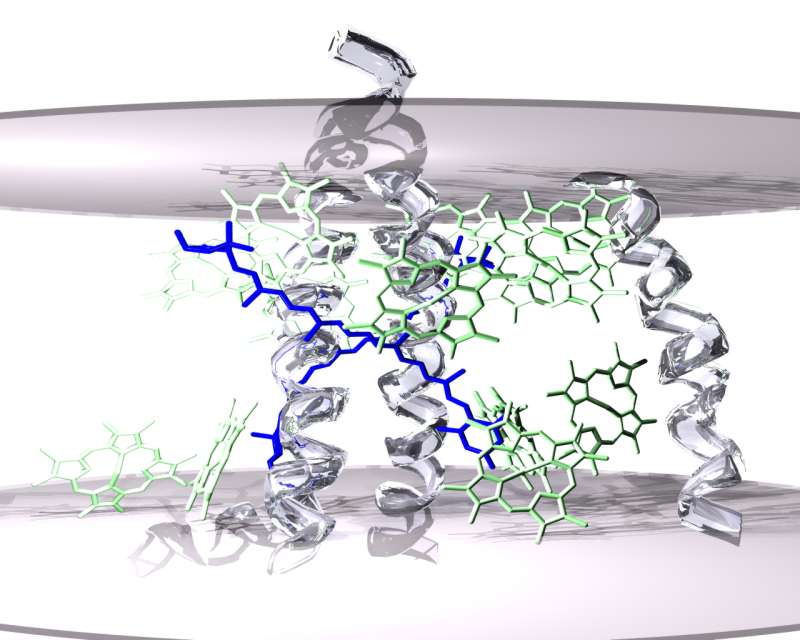 Figure B-10
Structure of LHC II inserted in the membrane (position of the
phospholipidic bilayer indicated by the grey discs).
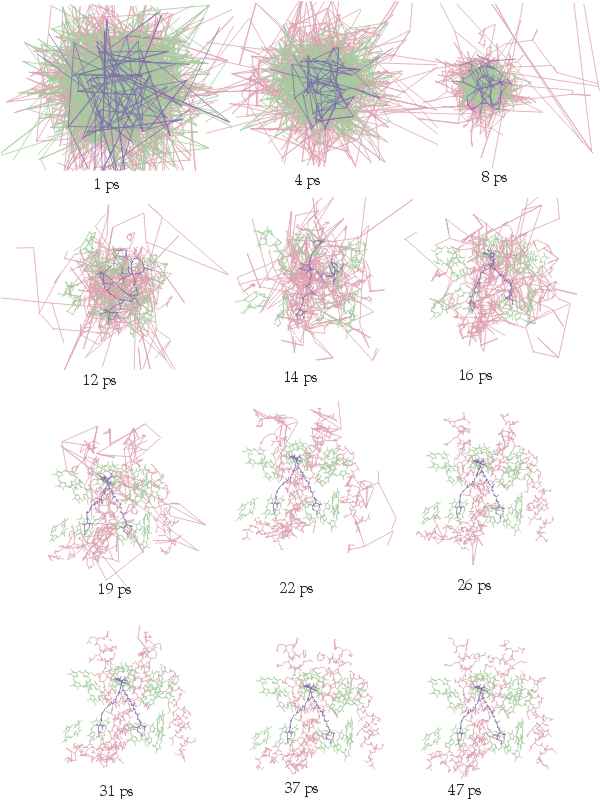 Figure D-19
Frames of the molecular simulation
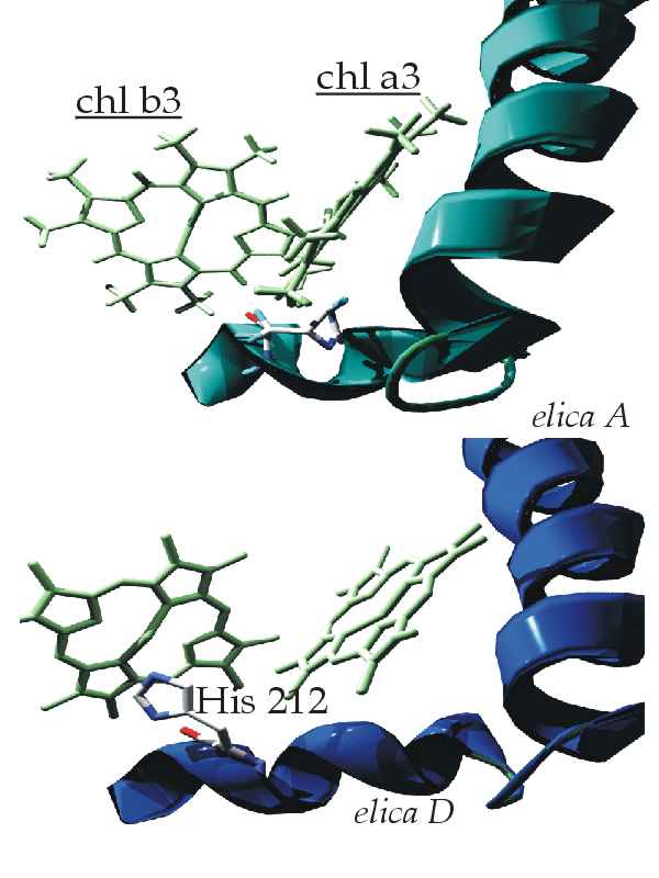 Figure D-20
Graphic representation of the simulation results. Residue His212 after
a normal minimization or normal simulated annealing (above). The same
residue after application of the protocol of simulated annealing using
relaxed force constants (below).
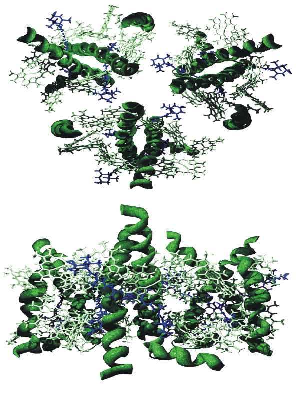 Figure D-31
The reconstructed trimer structure. Top view - Side view.
 Figure D-34
Hypothetical complex salt bridges
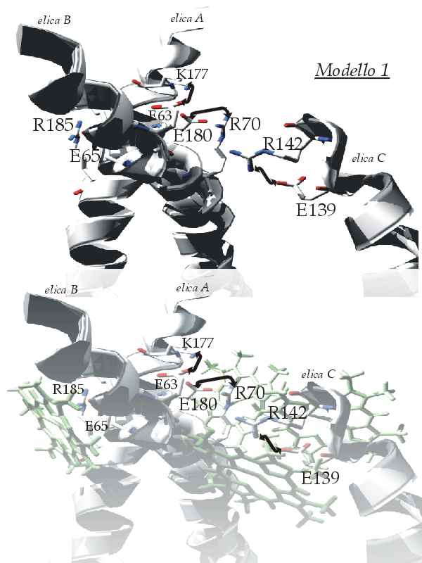 Figure D-35
Model 1. With (above) and without (below) the chlorophylls.
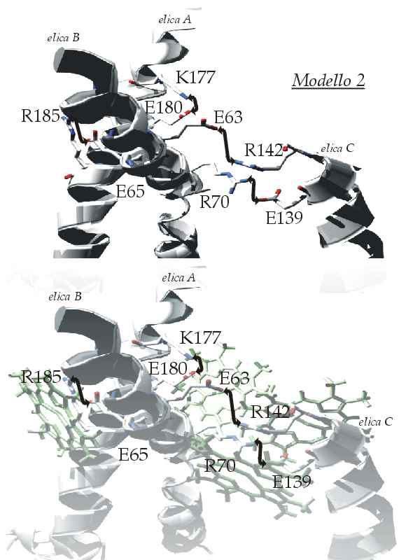 Figure D-36
Model 2. With (above) and without (below) the chlorophylls.
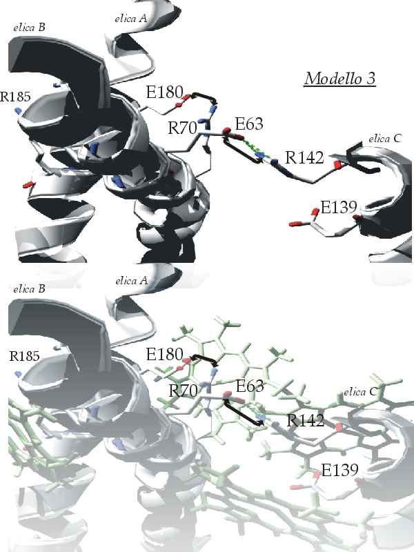 Figure D-37
Model 3. With (above) and without (below) the chlorophylls.
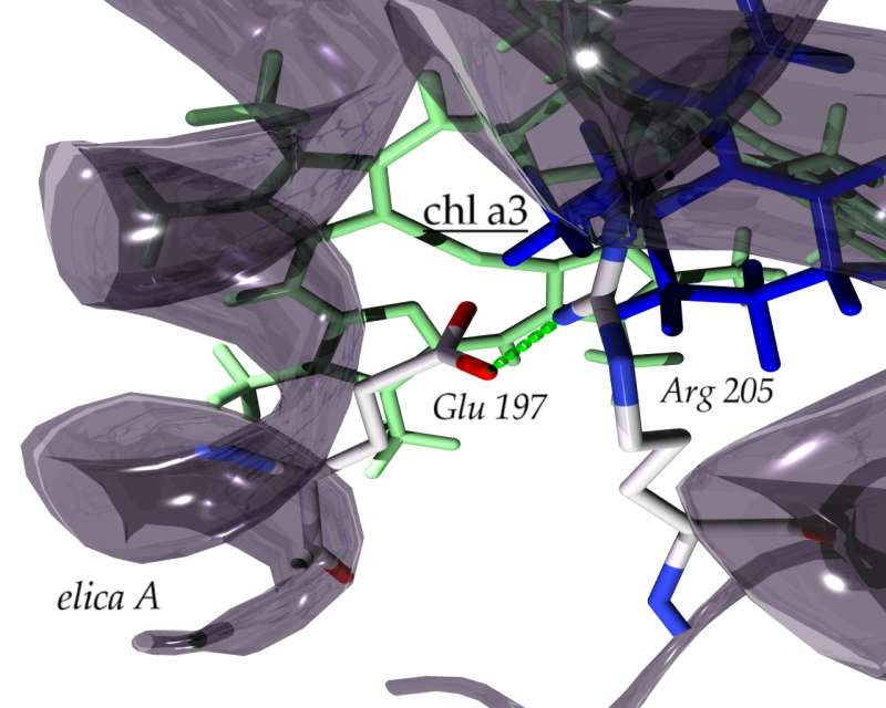 Figure D-38
The hyphotetical coordination of chlorophyll a3 in the CP24 protein